Key point The supraclavicular approach blocks the brachial plexus at its most compact point — the trunks/divisions — providing dense, reliable anaesthesia for elbow, forearm, and hand surgery with a single injection.
Anatomy
The brachial plexus is formed by the anterior rami of C5–T1. At the supraclavicular level, it has reorganised into three trunks (superior C5–6, middle C7, inferior C8–T1) dividing into anterior and posterior divisions. This is the most compact arrangement — ideal for complete block with a single injection.
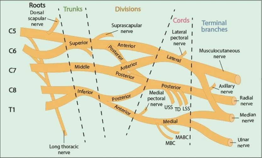
Key relationships
- The plexus lies lateral and posterior to the subclavian artery in the supraclavicular fossa
- Sandwiched between anterior and middle scalene muscles
- The first rib lies inferiorly — important depth landmark and safety floor
- The pleural dome is immediately posteromedial — pneumothorax is the classic complication
- The phrenic nerve (C3–5) runs on the anterior scalene and is frequently blocked (~50–80%)

Indications
Provides anaesthesia/analgesia for the elbow, forearm, wrist, and hand. For complete shoulder coverage, an interscalene block is preferred.
| Surgical Site | Block Role | Notes |
|---|---|---|
| Elbow surgery (distal humerus, olecranon, radial head) | First-line | Excellent coverage of all approaches |
| Forearm fractures, ORIF radius/ulna | First-line | Dense sensorimotor block |
| Wrist and hand surgery | First-line | May supplement with distal blocks for ulnar border |
| Carpal tunnel release | Alternative | Wrist blocks often preferred for simple CTS |
| Shoulder surgery | Partial | Prefer interscalene for complete shoulder coverage |
| AV fistula creation | First-line | Sympathectomy may improve vessel calibre |
Contraindications
Absolute
- Patient refusal
- Infection at injection site
- Allergy to local anaesthetics
Relative
- Contralateral pneumothorax or pneumonectomy
- Severe respiratory disease (FEV₁ <50%) — phrenic palsy risk
- Pre-existing coagulopathy
- Pre-existing neuropathy (document carefully)
Technique — Ultrasound-Guided
Real-time ultrasound guidance is strongly recommended to reduce the risk of pneumothorax and vascular injection. Nerve stimulation alone is no longer standard of care.

Equipment
- Probe: High-frequency linear (10–15 MHz), placed parallel to clavicle in supraclavicular fossa
- Needle: 50 mm short-bevel insulated 22G, in-plane approach from lateral
- Position: Supine, head turned 45° away, ipsilateral arm alongside body
Step-by-step
-
Initial scan — identify subclavian artery
Place probe in the supraclavicular fossa in coronal-oblique orientation. Identify the subclavian artery — pulsatile, compressible, anechoic circle. Confirm with colour Doppler if needed. Note depth of pleura.
-
Identify brachial plexus
The plexus appears lateral and slightly posterior/superior to the artery as a cluster of hypoechoic oval structures ("bunch of grapes"). The first rib is the bright hyperechoic band with posterior acoustic shadow beneath the artery.
-
Needle insertion — lateral to medial, in-plane
Insert the needle at the lateral end of the probe. Advance under real-time visualisation targeting the "corner pocket" — the space between the inferior trunk and the first rib, postero-inferior to the artery. This position uses gravity to distribute LA around all trunks.
-
Aspiration and incremental injection
Aspirate before each aliquot. Inject 5 mL increments with intermittent aspiration. Observe circumferential spread. Total volume 25–35 mL. Reposition needle if spread appears unilateral.
Sonoanatomy
On the US image the brachial plexus appears as a cluster of hypoechoic (dark) oval/round structures lateral to the subclavian artery. The first rib produces a bright white hyperechoic band with posterior acoustic shadow. The pleura appears as a thin, bright, sliding line posteromedial to the artery.
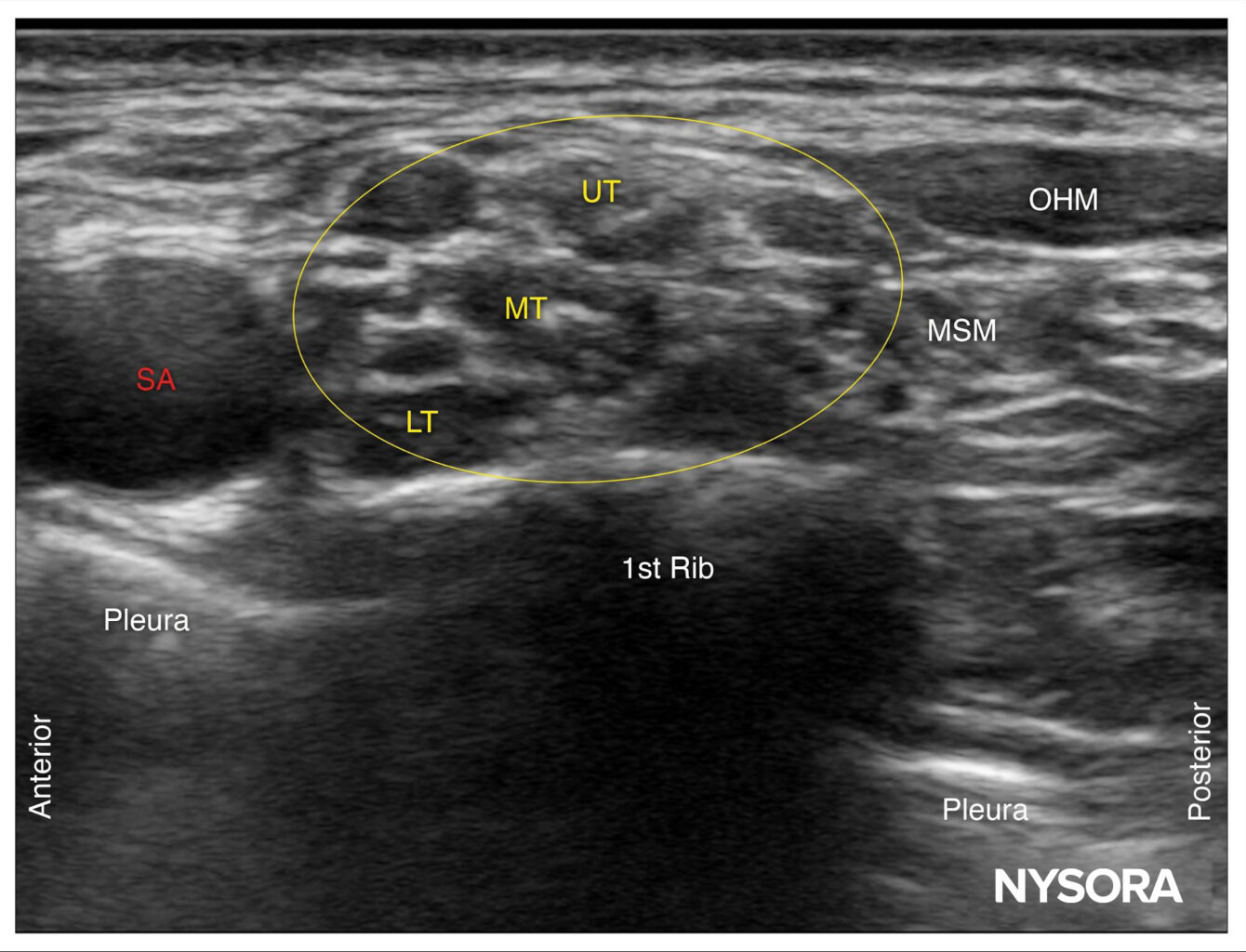
Image courtesy of NYSORA (nysora.com). Source: Tips for a Supraclavicular Brachial Plexus Block — NYSORA. Used for educational purposes.
Sonoanatomy tip: Start by locating the subclavian artery — pulsatile dark circle. Then look laterally and slightly posteriorly for the plexus cluster. The first rib produces a bright white line with complete posterior shadowing. The "corner pocket" injection point is the angle between the inferior plexus and the first rib, posteromedial to the artery.
Local Anaesthetics & Dosing
Maximum dose limits must always be respected. Calculate mg/kg dose before injection. Consider dose reduction in elderly, low weight, or hepatic insufficiency. Verify against local formulary and current SmPC before clinical use.
| Agent | Concentration | Volume | Dose | Onset | Duration (plain) | +Adrenaline |
|---|---|---|---|---|---|---|
| Ropivacaine | 0.5% | 25–35 mL | 125–175 mg | 15–25 min | 8–12 h | — |
| Ropivacaine | 0.75% | 20–30 mL | 150–225 mg | 10–20 min | 10–16 h | — |
| Bupivacaine | 0.5% | 20–30 mL | 100–150 mg | 15–25 min | 10–16 h | 14–20 h |
| Levobupivacaine | 0.5% | 20–30 mL | 100–150 mg | 15–25 min | 10–14 h | 12–18 h |
| Lidocaine | 1.5–2% | 20–30 mL | 300–400 mg | 5–10 min | 2–4 h | 3–6 h |
| Mepivacaine | 1.5% | 25–35 mL | 375–525 mg | 8–15 min | 4–6 h | 6–8 h |
Ropivacaine 0.5% — 30 mL (150 mg total)
+ Dexamethasone 8 mg IV as adjuvant. Provides 12–16 h analgesia in most patients.
Mepivacaine 1.5% 20 mL + Ropivacaine 0.5% 15 mL (mixture)
Fast onset ~10 min, extended duration ~8 h. Useful when rapid surgical onset is required.
Adjuvants
| Adjuvant | Dose | Route | Effect | Evidence |
|---|---|---|---|---|
| Dexamethasone | 4–8 mg | IV or perineural | Extends duration by 6–8 h, ↓ opioid use | Strong |
| Dexmedetomidine | 0.5–1 mcg/kg | Perineural or IV | Extends duration by 4–6 h | Moderate |
| Adrenaline | 2.5–5 mcg/mL | With LA | Extends duration (lidocaine/mepivacaine); intravascular marker | Strong |
| Clonidine | 150 mcg | Perineural | Modest extension; sedation possible | Limited |
Complications
| Complication | Incidence | Risk | Mitigation |
|---|---|---|---|
| Phrenic nerve palsy | 50–80% | High | Reduced volume (15–20 mL); counsel patients with respiratory compromise |
| Pneumothorax | 0.1–0.7% (US-guided) | Moderate | Identify pleura before injection; real-time US; in-plane technique |
| Vascular puncture | 1–3% | Moderate | Colour Doppler; in-plane visualisation; avoid medial trajectory |
| LAST | <0.1% | Low | Incremental injection; aspirate; Intralipid 20% immediately available |
| Horner syndrome | 1–5% | Low | Self-limiting; warn patients preoperatively |
| Persistent neuropathy | <0.05% | Low | Avoid intraneural injection; low injection pressure; document pre-existing deficits |
Intralipid 20% (500 mL) must be immediately available whenever performing any peripheral nerve block. Ensure full LAST resuscitation kit is present. Follow AAGBI LAST guidelines.
Video Resources
Educational videos embedded from YouTube. All content is from educational/institutional channels.
Supraclavicular Block — Anatomy & Technique
Anatomy, indications, and ultrasound-guided technique walkthrough.
Scanning Technique & Sonoanatomy
Detailed review of US scanning technique and sonographic landmarks.
Finding the Brachial Plexus — US Landmarks
Step-by-step probe placement and landmark identification for beginners and intermediates.
Live Procedure Demonstration
Real-time US-guided supraclavicular block demonstration with commentary.
References
- Tran DQH et al. Supraclavicular nerve blocks: systematic review and meta-analysis. Reg Anesth Pain Med. 2017;42(5):556–566.
- Neal JM et al. ASRA Practice Advisory on Local Anesthetic Systemic Toxicity. Reg Anesth Pain Med. 2018;43(2):113–123.
- NYSORA. Ultrasound-Guided Supraclavicular Brachial Plexus Block. nysora.com ↗
- ESRA PROSPECT Working Group. Procedure-specific postoperative pain management recommendations. esraeurope.org/prospect ↗
- NYSORA. Tips for a Supraclavicular Brachial Plexus Block. nysora.com ↗ [Ultrasound image, used for educational purposes]
- Abdallah FW, Brull R. Perineural dexmedetomidine: systematic review. Br J Anaesth. 2013;110(6):915–925.
- Desmet M et al. Intravenous dexamethasone and nerve block duration. Br J Anaesth. 2017;117(6):799–807.
- King of the Curve. Understanding Brachial Plexus Injury: Nerve Anatomy and Clinical Relevance. kingofthecurve.org ↗ [Anatomical diagram, used for educational purposes]